BLOOD, CUTANEOUS, AND PULMONARY PARASITE
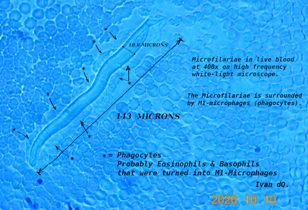
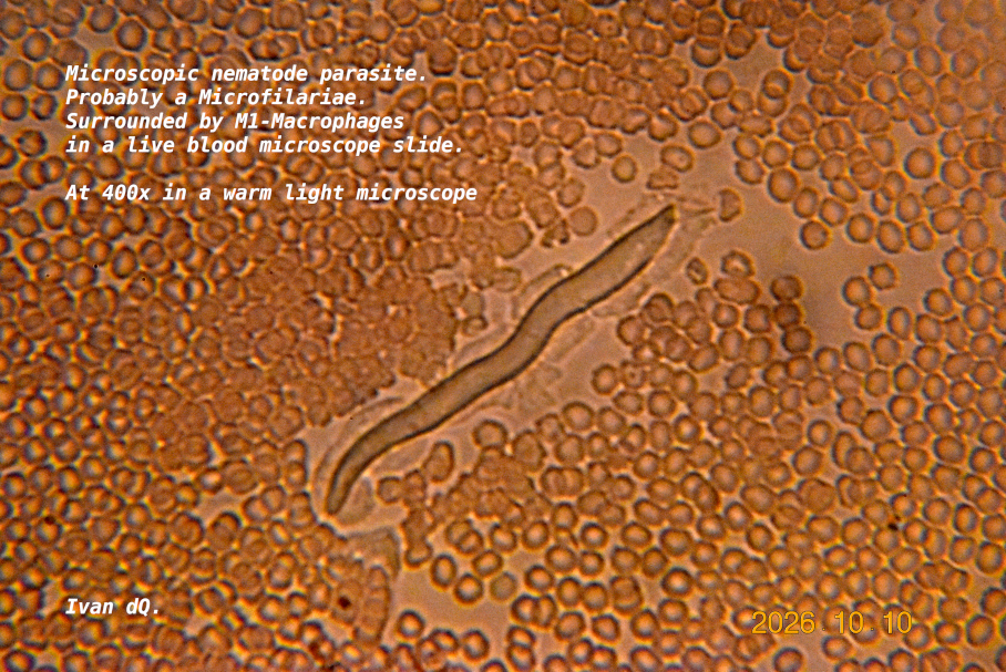
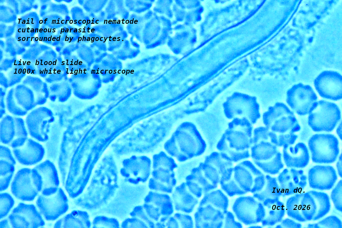
On October 10, 2026, I made a microscopic slide from a tiny drop of
blood drawn from the skin, of my arm, in an area that had been
itching for a few days and had tiny skin nodules. I patiently
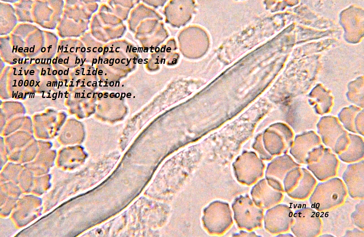
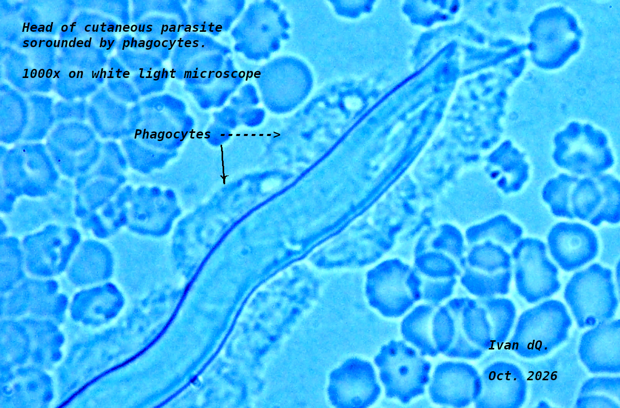
scanned the whole slide under my microscopes. And I found a
microscopic nematode (worm) that was being attacked and surrounded
by numerous phagocytes.
On September, a blood test had shown that the Eosinophils and the
Basophils, in my blood, were higher than normal. This usually
indicates a parasitic infection. Therefore, I believe that the
phagocytes, that were surrounding the microscopic nematode, were
probably Basophils and Eosinophils that had been activated as
M1-macrophages.
I was able to capture some really good micrographs of the micro
nematode. And I measured the size of it using the Pixels to Microns Conversion Table
that can be found in my Microscopy portal.
This parasite could be a baby microfilariae, like larger versions
that I have previously seen in my sputum and blood.
The micrographs were captured using a Nikon D3500 digital camera
with a 10x microscope lense that fits in the triport or the eyepiece
tube of the ocular lense. The blueish micrographs were taken using
an LED white light microscope. The others were taken with a warm
light microscope.
So, I am including, in this web page, some of the micrographs that I
took.
Select (mouse click) on the small images bellow to see the larger
micrographs.


RETURN TO
MICROSCOPY PORTAL
RETURN
TO HEALTH MATTERS PORTAL
RETURN TO MANY
PORTALS