BLACK AND WHITE MICRO-VIDEOS OF PHAGOCYTES (M1 MACROPHAGES)
I am neither a hematologist, nor a microbiologist, nor do I know all
of the medical studies required to be a hematologist or a
microbiologist. However, I consider myself an amateur microscopist
who is very curious about what is in my blood. And I have been able
to capture micro-graphs and micro-videos that clearly show the
different blood cells, some stained and some alive and fully
functional, such as some phagocytes (M1 macrophages) cleaning the
the blood from bacteria and microscopic parasites.
Those persons, studying any medical science, or curious about blood
cells and pathogens found in the blood, might find these
micro-graphs and micro-videos interesting.
Blood Phagocytes can be monocytes, macrophages, neutrophils, or mast
cells.
Phagocytes are also called M1 Macrophages.
Information, other than the micro-graphs and micro-videos, are
backed by reference links to Wikipedia or medical text books. I try
my best to not spread misinformation or disinformation.
In 2013, I only had an inexpensive microscope digital camera that
could only take black and white videos in AVI or WMV formats. Bellow
are four of those videos. The ones in AVI formats run faster than
normal speed.
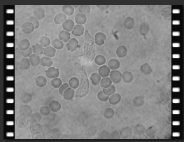
These many busy phagocytes in a live blood slide is usually
indicative of an infection.
51 sec in avi format
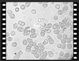
The black & white micro-video bellow shows phagocytosis
occurring. Notice near the center of the micro-video, what looks
like a small bright circle or bubble at the end of a phagocyte,
breaks open, releasing the debris of the pathogen that was destroyed
inside the phagosome. However, along with such debris, superoxydants
(that can cause inflammation) are also released. I tried to shorten
the 2.2 minutes video, but my video editing software programs (3 of
them) do not like wmv format (Windows Multimedia Video Format).
2.2 minutes in wmv format
The two micro-videos bellow show what seems like infected phagocytes
or eosinophils in their phagocytosis mode. There are certain
pathogens that, when engulfed by a macrophage-phagocite, they avoid
getting trapped in a phagosome. Then they can reproduce inside the
macrophage, eventually rupturing the macrophage and going on to
infect other cells.


3 seconds avi format
16 seconds avi format
REFERENCES:
https://en.wikipedia.org/wiki/Phagocyte
https://en.wikipedia.org/wiki/Phagocytosis
https://en.wikipedia.org/wiki/Phagosome
https://en.wikipedia.org/wiki/Macrophage
https://en.wikipedia.org/wiki/Neutrophil
https://en.wikipedia.org/wiki/Plasmodium
RETURN