FAILURE TO FOLLOW THE CUSTOMARY STANDARD OF
SAVING THE DICOM IMAGES, OF A CT SCAN, IN THE PROPER SEQUENCE.
Before I describe the malicious scrambling of the
tomographic images of CT scans, that is done by new Windows software
(from Altamond Software Inc) being included in some CT scan DVDs
given to patients of the Miami VA Health Care System. I must include
a brief introduction and description of CT scans and the DICOM
format used to store the tomographic images created during a CT
scan.
A Computed Tomography Scan (CT scan) formerly known as Computed
Axial Tomography Scan (CAT scan) is a medical imaging technique used
to obtain detailed internal imaging of the body. In my humble
opinion, they should not have removed the "axial" word from it's
name, because it is very important to know that the tomography
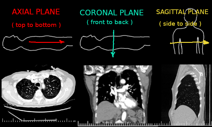
images, that look like X-ray slices, are captured sequentially in 3
different axis (planes).
The three planes (axis), used to capture the tomography images
(x-ray slices), are named:
Sagittal plane, Coronal plane, and Axial plane.
Bellow I show the direction used to capture (sequentially) the
tomography images in each of the defined planes. And I include a
sample image of each.
Each image (X-ray slice) is saved, in a sequentially numbered
computer file, in a unique format called DICOM (Digital Imaging and
COmmunications in Medicine). The sequentially numbered files are all
stored in a folder (directory) named DICOM. Sometimes the images of
each plane are stored (sequentially numbered) in separate
sub-folders named Series-nn .The National Electrical Manufacturers
Association (NEMA) holds the copyrights to the DICOM standard and
they make it available for download, from their website, for free,
in many formats (i.e. PDF, HTML, etc). However, I believe that the
standard does not specifies that the DICOM images should be store
sequentially in the media that is given to patients. Probably
because it is obvious, logical, and rational to do so. Only an idiot
or a malicious software developer would scramble the DICOM images
files of all three planes, and renumber the DICOM file names with
numbers different than the numbers that were assigned by the CT
scanner when the images where created.
Why would a malicious software developer scramble and renumber the
DICOM images of all three planes? To ensure that only their
Windows software will be able to display the DICOM images
sequentially. Anyone trying to view the images, with a different
Windows application (DICOM viewer), or trying to view them one at a
time, sequentially, using a different viewer, from a different OS,
would not be able to do so.
The only viewer that does this crazy scrambling and false number
renaming of files, in the DICOM folder (directory), is the new
viewer that the Miami VA is including in their CT-scan DVDs. The
viewer is made by "Altamont Software". Shame on you Altamont
Software.
The VA is also using "ClearRead CT" from Riverain
Technologies. An excellent AI DICOM image analyser, capable of
finding and measuring anomalies in DICOM images and doing a
differential diagnosis. Kudos to Riverain Technologies developers
for creating such an excellent software.
However, for me to find the ClearRead CT summary files in the DICOM
folder, I had to open each of the 799 scrambled and renumbered DICOM
files, in the DICOM directory. So that I could print the ClearRead
Summaries. And that took me longer than the time I spent creating
this HTML file for this portal.
The Miami VA previously used the "Centricity DICOM viewer" later
replaced by "Gearview". Those viewers did not scramble the DICOMs.
Niether does other viewers such as pbcdview, Viewlite, and Rediant.
UPDATE (August 31):
What a weird coincidence! The morning after I first posted
this page, an administrator from the radiology department of the
Miami VA, called me on the phone. About some CT scan survey that I
had responded to a few days ago. However, the administrator did not
have the necessary technical knowledge to understand the problems
created by scrambling and renumbering the files in the DICOM folder
(directory). One of the questions he asked was "How is it you know
these things?", and he is wright, must people (perhaps 99.99 %) do
not view DICOM images individually, with special viewers, nor do
they colorized the images. But I wonder "How is it that he does
not?". I will try to send him a link to this page, so that he
becomes more knowledgeable regarding DICOM images.
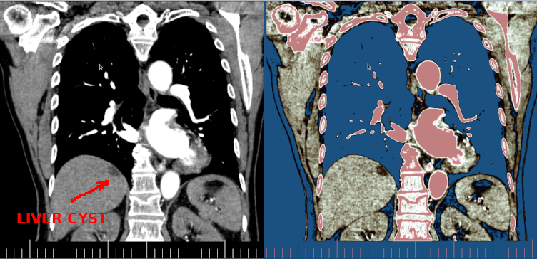
I am guessing that he has never viewed a DICOM image with a good
DICOM (single file viewer) that allows the user to shift the grey
scales used in the DICOM images and also change the contrast of the
DICOM images. I can even "colorize" the images, I am
including a colorized image bellow to illustrate how a cyst in a
liver is more visible when the image is colorized.
REFERENCE LINKS:
https://en.wikipedia.org/wiki/CT_scan
https://commons.wikimedia.org/wiki/Scrollable_high-resolution_computed_tomography_images_of_a_normal_thorax
RETURN